

The trade-off between radiation exposure and image quality

Many people fear LCS may be an invasive procedure, could lead to incidental findings, or involve radiation exposure.1
To learn more about what stops people from participating in LCS, check out the first edition in our series
In diagnostic imaging, reducing patient dose often comes with the trade-offs of poorer image quality2,3 and increased clinical risk of patient mismanagement.3
The target for optimization is the point where the combined net risk is minimized.3
To deliver the benefits of LCS and minimize the effective dose (ED), many countries have provided recommendations4-8 on radiation dose.
Despite the guidelines, a wide distribution of
LCS CT doses across facilities continues to be reported.4
LCS CT doses across facilities continues to be reported.4
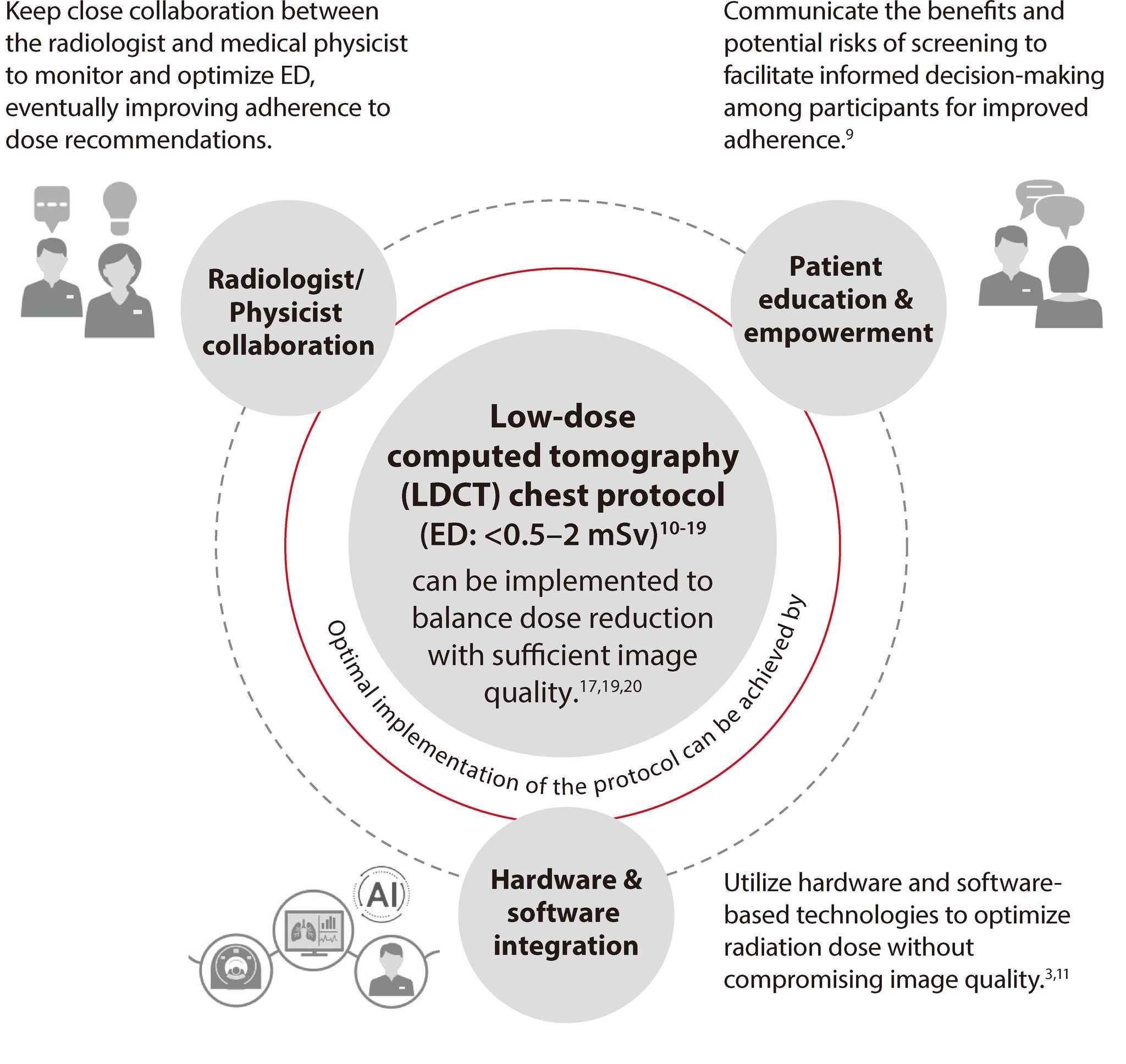
Strategies for reducing radiation dose while preserving image quality

Effective screening
program for asymptomatic
at-risk individuals
96%
of low-dose chest CT scans (0.44 mSv)
were reported to be of diagnostic
quality in a recent study.17
were reported to be of diagnostic
quality in a recent study.17
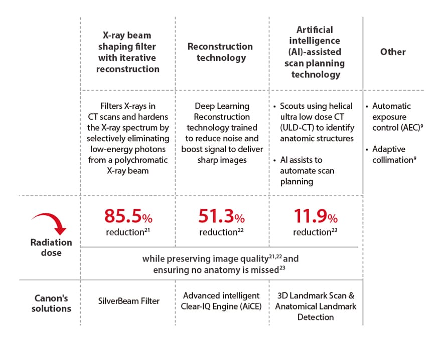
Hardware and software that can help you optimize the implementation of the LDCT protocol
Examples & range of dose reductions based on data from recent studies:
Subscribe and Stay Updated
Subscribe to our newsletter to receive campaign updates, access to online webinars and other educational information.
- References:
References: - 1. Cavers D, et al. Respir Res. 2022;23(1):374. 2. Barrett HH, et al. Phys Med Biol. 2015;60(2):R1-R75. 3. ICRP PUBLICATION 154. Ann ICRP. 2023;52(3):11-145. 4. Demb J, et al. JAMA Intern Med. 2019;179(12):1650-1657. 5. Pereira LF, et al. J. Bras. Pneumol. 2024;50(01):e20230233. 6. Snoeckx A, et al. Transl Lung Cancer Res. 2021;10(5):2356-2367. 7. ESTI LCS technical standards. 2019. Available from: https://www.myesti.org/content-esti/uploads/ESTI-LCS-technical-standards_2019-06-14.pdf. Accessed October 18, 2024. 8. China Lung Oncology Group. Chin J Lung Cancer. 2023;26(1):1-9. 9. Dudhe SS, et al. Cureus. 2024;16(5). 10. National Lung Screening Trial Research Team. N Engl J Med. 2011;365(5):395-409. 11. Pozzessere C, et al. Tomography. 2023;9(1):166-177. 12. Larke FJ, et al. AJR Am J Roentgenol. 2011;197(5):1165-1169. 13. Macri F, et al. Diagn Interv Imaging. 2016;97(11):1131-1140. 14. Gobi K, et al. Pol J Radiol. 2022;87:e597-e605. 15. Paks M, et al. Medicine (Baltimore). 2018;97(34):e12019. 16. Ludwig M, et al. BMJ Open. 2019;9(8):e025661. 17. Kim Y, et al. AJR Am J Roentgenol. 2015;204(6):1197-1202. 18. Newell Jr JD, et al. Invest Radiol. 2015;50(1):40-45. 19. Kroft LJ, et al. J Thorac Imaging. 2019;34(3):179-186. 20. Greffier J, et al. Diagn Interv Imaging. 2020;101(6):373-38. 21. Golbus AE, et al. Eur Radiol. 2024;34(9):5613-5620. 22. McLeavy CM, et al. UKIO Online. 2020;P094:5. 23. Golbus AE, et al. Eur J Radiol Open. 2024;13:100578.
Social Media


