

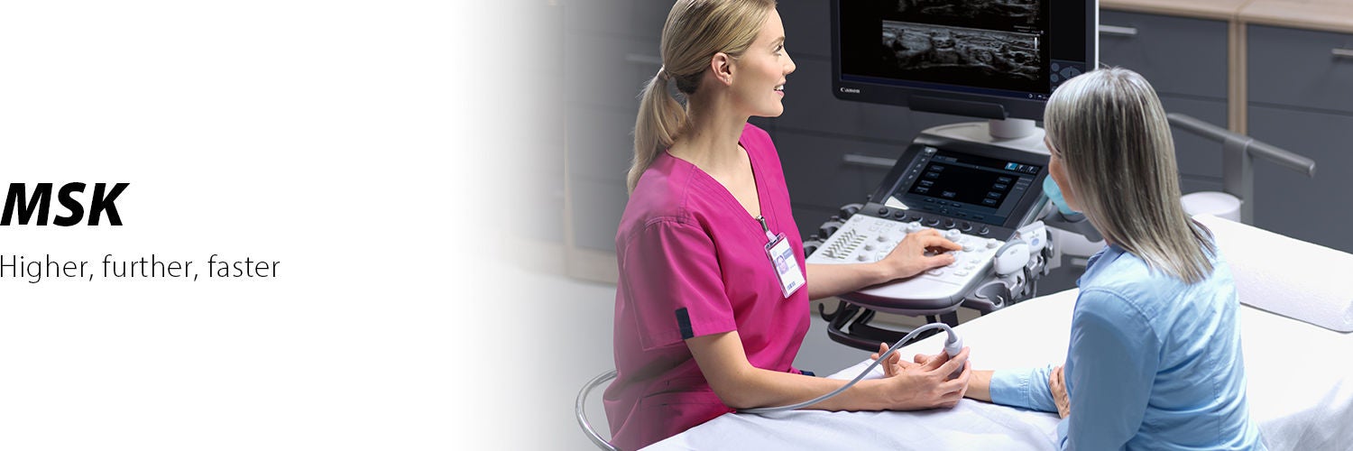
Musculoskeletal clinical solution
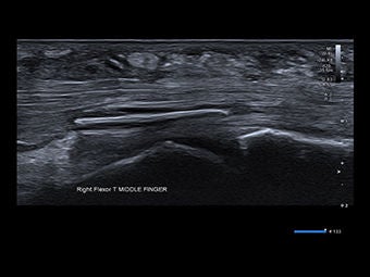
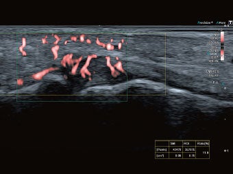
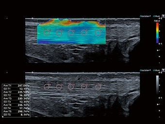
Musculoskeletal (MSK) imaging is moving beyond muscles and tendons. For example, identifying the exact location of very small foreign bodies close to moving structures of the MSK anatomy is important when planning best method of removal. Another indicator of pathology in MSK studies is the presence of increased blood flow due to inflammation. Accurate detection of increased blood flow can help in monitoring the effect of therapy and in patient management.
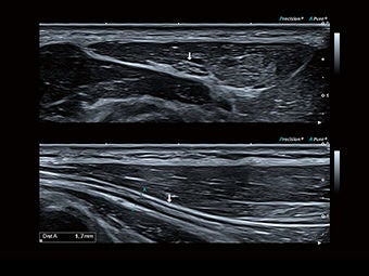
Canon’s improved matrix technology, intelligent Dynamic Micro-Slice (iDMS) allows more control over the beam to provide a super thin slice thickness in both planes for outstanding high resolution imaging, in focus all the way from the near to far field. This new generation of matrix transducers is unique to Canon and an advancement on already existing matrix technology that we have utilized for many years.
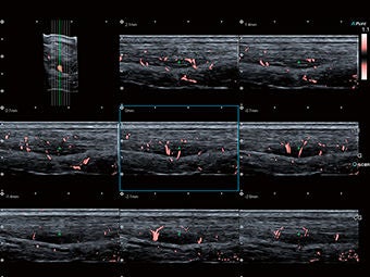
Our revolutionary Supeb Micro-vascular Imaging (SMI) allows the visualization of the micro-vascularity of MSK structures which can be impossible to detect with conventional color Doppler. SMI provides more clinically useful information in MSK examinations and has set a new standard in detecting micro-vascularity.
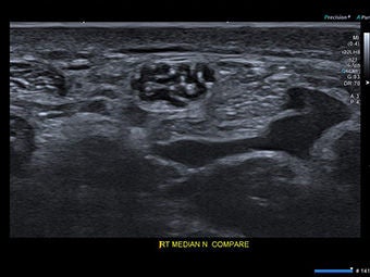
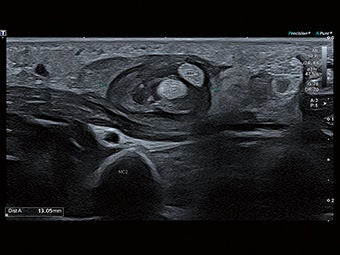
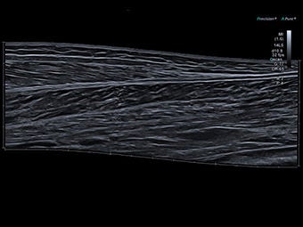
Canon continues this revolution with the introduction of the 33MHz transducer. This high frequency transducer is able to demonstrate detail in the skin layers, as well as superficial ligaments and tendons that has up until now been impossible to see.
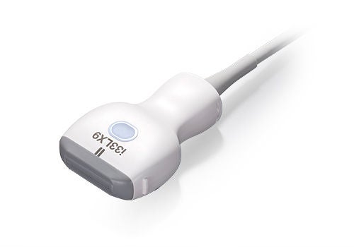
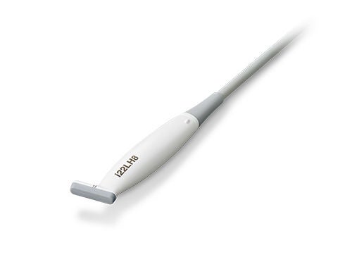
High resolution in a small footprint for sub-millimeter imaging
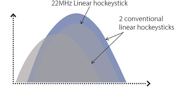
22MHz High frequency hockeystick transducer
Aplio’s 22MHz hockeystick provides the high frequency needed for the most demanding small parts examinations.
New features:
- Very high resolution
- Ergonomic design with small 8mm footprint and slim cable, great for examining collateral ligaments and other finger and hand pathology
- SMI-capable
Clinical images
Education and resources





White paper

Corinne E. Wessner BS, RDMS, RVT,
Priscilla Machado MD, Shawn Peterson DO,
Flemming Forsberg PhD, Kibo Nam PhD
High Frequency Ultrasound Imaging for Diagnosing Carpal Tunnel Syndrome
Akihiro Narita, MD, PhD
Norifumi Sawamukai, MD, PhD
Clinical value of Superb Micro-vascular Imaging and ultra-high frequency linear transducer in musculoskeletal ultrasound:
- From early detection to diagnosis and treatment evaluation -

Professor Adrian K.P. Lim
Preliminary experience of ultra-high frequency imaging with the Aplio i-series
Dr Steve McNally
Superb Micro-Vascular Imaging – A new tool for the sports physician
Interview report & Case study
Seeking more information? We can help.

Social Media


