Search
Region / Language
Contact Us
Products & Solutions
Products
Angiography
Products
Alphenix
Alphenix / Evolve Edition
Alphenix 4D CT - Angio-CT suite
Clinical segments
Interventional Cardiology
Interventional Radiology / Oncology, Vascular
Interventional Neurology
Computed Tomography
Area Detector CT
Aquilion ONE / INSIGHT Edition
Aquilion ONE / PRISM Edition
Multi Slice CT
Aquilion Serve SP
Aquilion Serve
Aquilion Prime SP
Aquilion Lightning 80
Aquilion Lightning
Aquilion Start
Large Bore CT
Aquilion Exceed LB
Other products
Photon Counting CT
Mobile CT
Technologies
Advanced intelligent Clear-IQ Engine (AiCE)
Precise IQ Engine (PIQE)
Deep Learning Spectral
SURE Subtraction
AIDR3D
More information
CT Dynamic Volume MSK
Magnetic Resonance
3T MRI
Vantage Galan 3T / Supreme Edition
1.5T MRI
Vantage Orian
Vantage Fortian
Vantage Elan
Encore Upgrade
Vantage Orian / Encore Upgrade
Technologies
Advanced intelligent Clear-IQ Engine (AiCE)
Precise IQ Engine (PIQE)
More information
Good to know
Download Protocols
Ultrasound
Clinical solutions
General Imaging
Womens' Health
Cardiovascular
Products
Aplio i-series / Prism Edition
Aplio beyond
Aplio a-series
Aplio me
Aplio flex
Aplio go
Aplio i700 - Women's Health
Aplio beyond - Women's Health
Aplio me- Women's Health
Technologies
3rd Harmonic Imaging
Differential Tissue Harmonic Imaging (D-THI)
Ultra Wide View
More information
Remote connection
X-ray
Fluoroscopy
Ultimax-i
Zexira i9
Raffine-i
Intelli-C
Radiography
Radrex i5 / Flex Edition
RADREX
Radrex Drite
CXDI-702C Wireless
CXDI-710C Wireless
Mobile X-ray
Mobirex i9 / Smart Edition
Mobirex i9
Specialized X-ray
NRT Products
Mammography
HIT
AI solution
Automation Platform
Advanced Visualization
Vitrea
Modality operation
Vina Analytics
Clinical Solutions
Stroke
Oncology
Lung Cancer Screening
SportsMed
Cardiology
Prostate
Innovations
AI
Efficient Workflow
Collaborative imaging
Service & Support
Service
Our Service
Service Vision
Service Solution
Remote Service
Refurbishment
Training and Education
Interoperability
Interoperability Index
DICOM Current Products
DICOM Past Products
IHE Current Products
IHE Past Products
Online Digital Manuals
Product Security Information
Product Security Information
Product Security Policy
Security Advisory
Product Security Inquiries
Product Security Update
Knowledge center
Canon Medical Academy
Publications
Visions Special
Angio Visions magazine
MRI Visions magazine
AI Visions magazine
Events
Events
About Us
Corporate Information
Sustainability Solutions
Made for Life
Made Possible
HR
Photo Gallery
×
Search
keyword
search
Aplio go
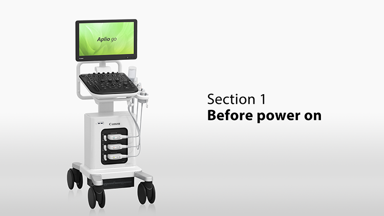
Section 1: Before power on
Watch Video
×
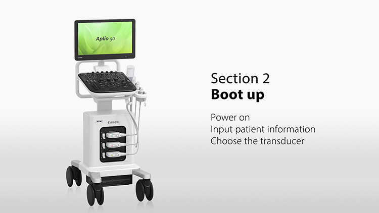
Section 2: Boot up
Watch Video
×
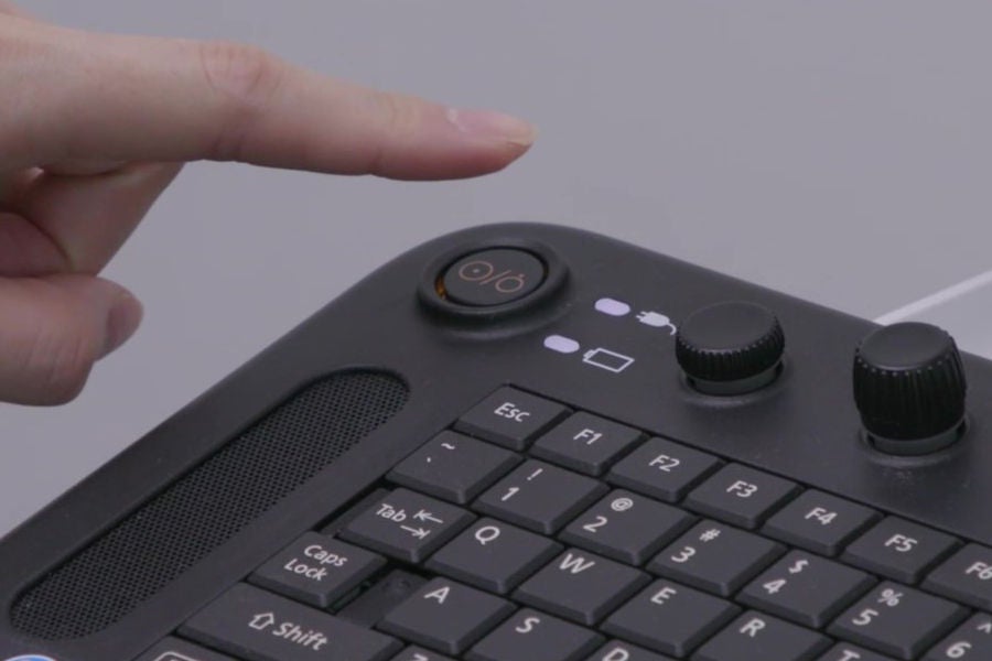
power on
Watch Video
×
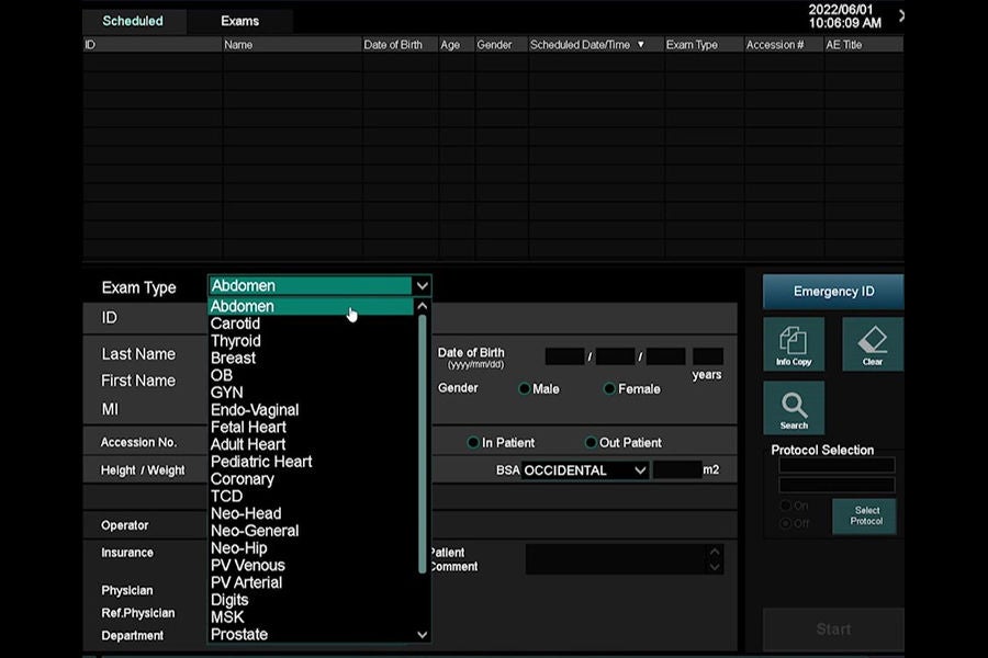
Enter patient information
Watch Video
×
Select the transducer
Watch Video
×
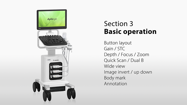
Section 3: Basic operation
Watch Video
×
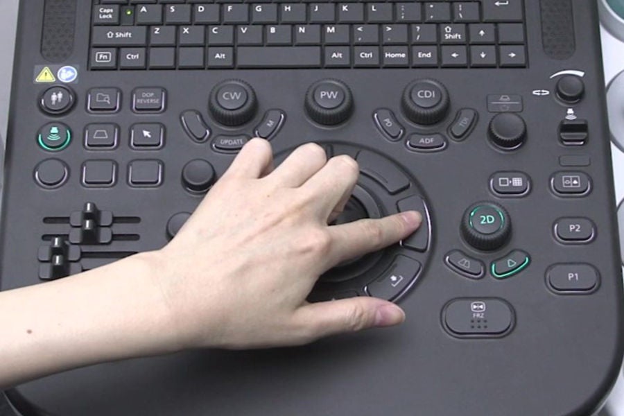
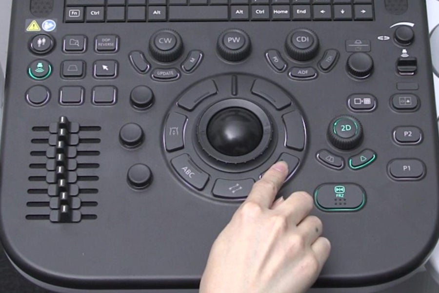
Button layout
Watch Video
×
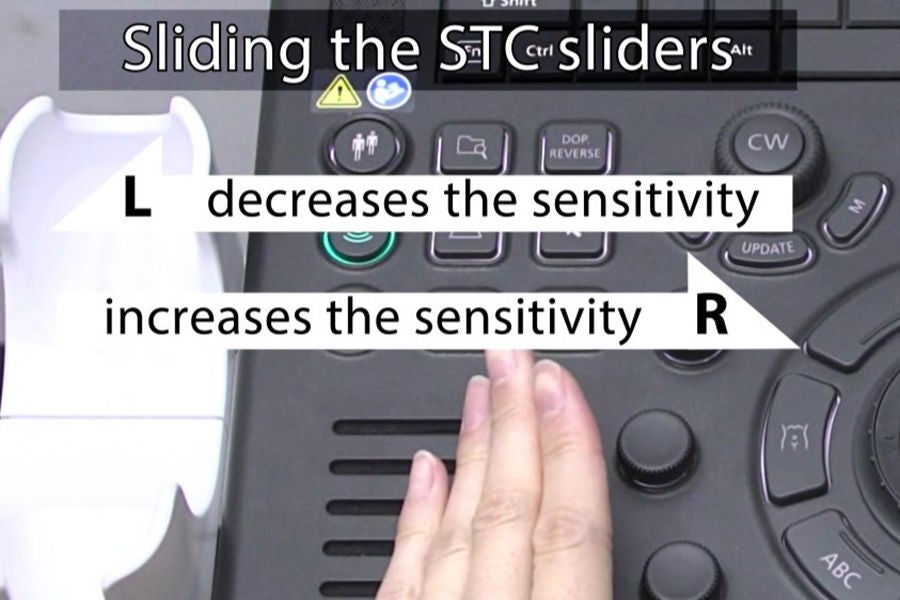
Gain / STC
Watch Video
×
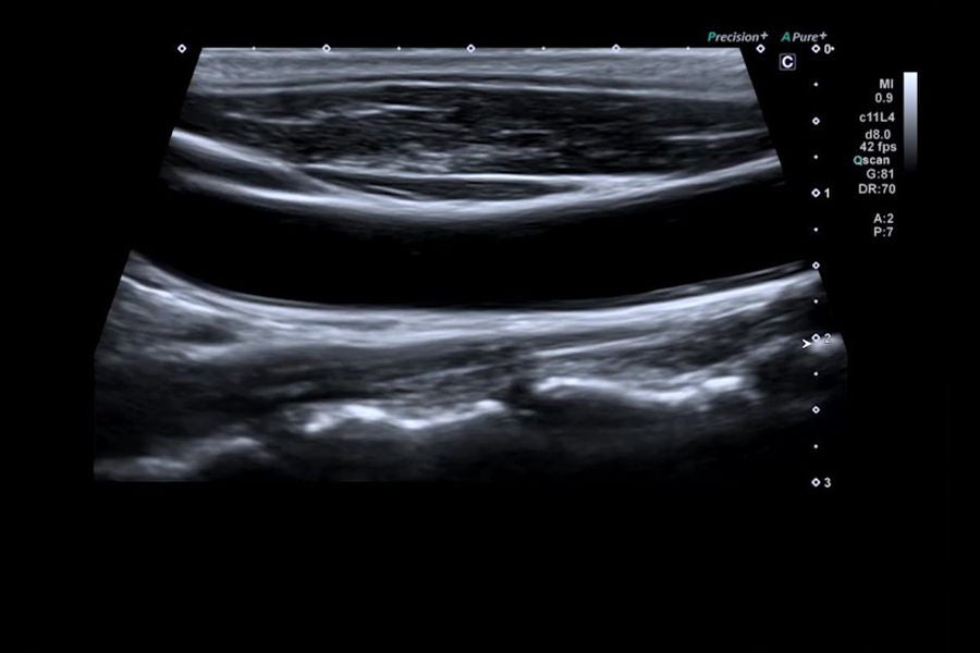
Depth / Focus / Zoom
Watch Video
×
Quick Scan / Dual B
Watch Video
×
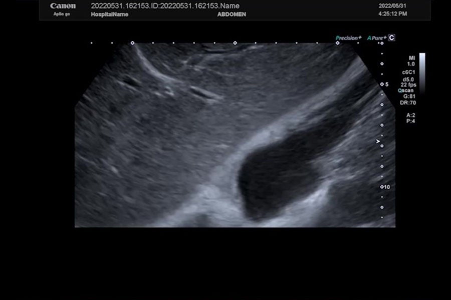
Wide view
Watch Video
×
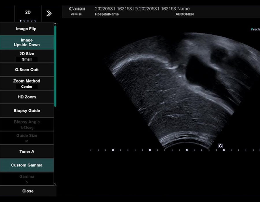
Image invert / up down
Watch Video
×
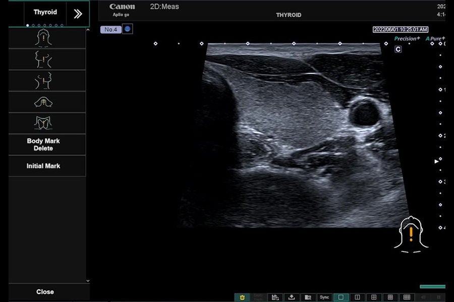
Body mark
Watch Video
×
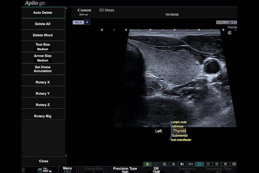
Annotation
Watch Video
×
Section 4: Color / Pulse doppler operation
Watch Video
×
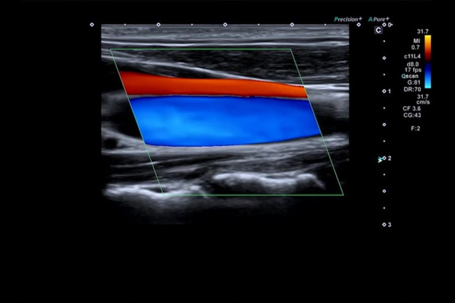
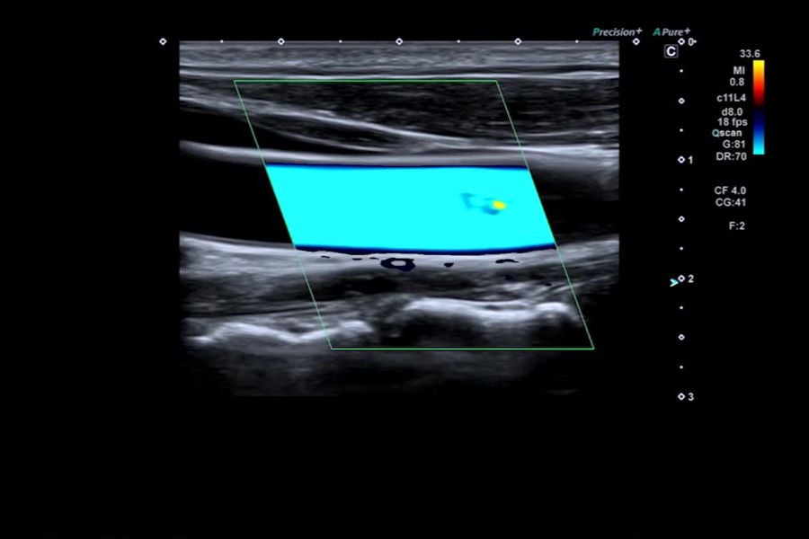
Color Doppler Imaging (CDI)
Watch Video
×
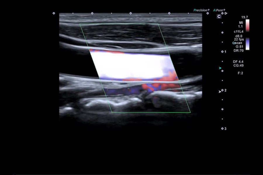
Power Doppler (PD)
Watch Video
×
Advanced Dynamic Flow (ADF)
Watch Video
×
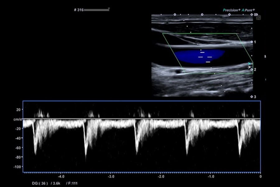
Pulse Wave Doppler (PW)
Watch Video
×
Section 5: Cardiac basic operation
Watch Video
×
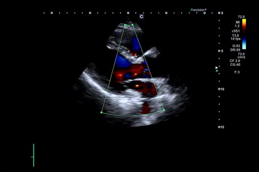
Color Doppler Imaging (CDI) for Cardiac
Watch Video
×
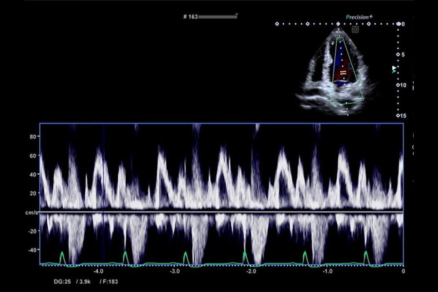
Pulse Wave Doppler (PW) for Cardiac
Watch Video
×
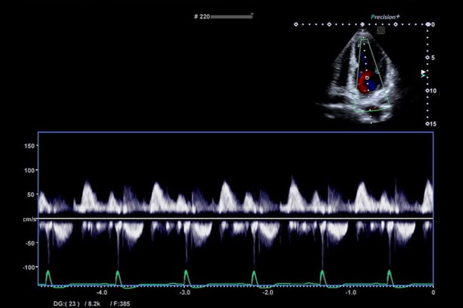
Continuous Wave Doppler (CW) for Cardiac
Watch Video
×
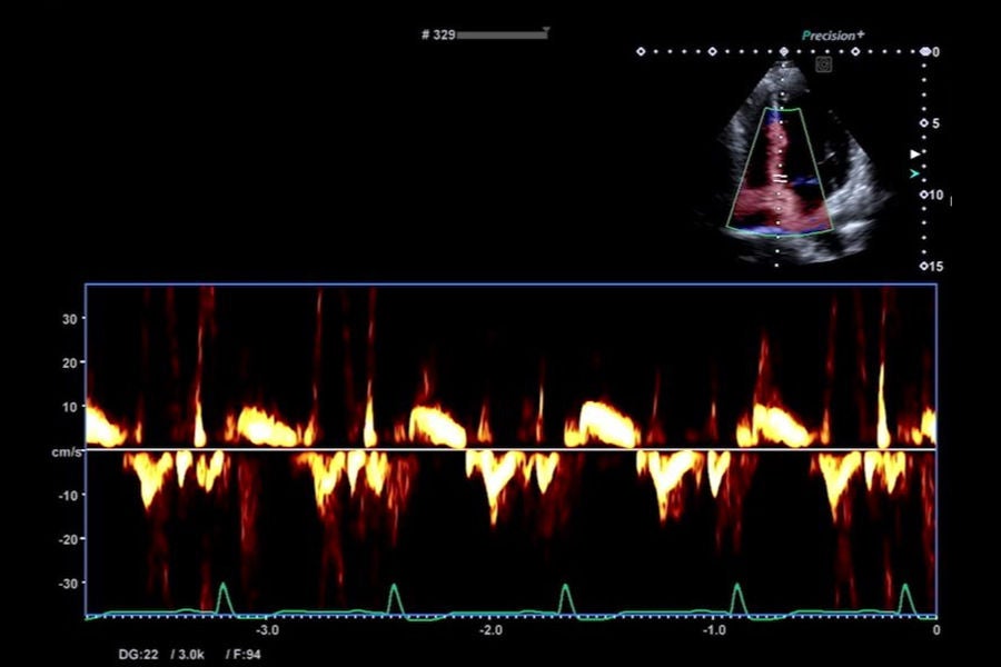
Tissue Doppler Imaging (TDI) for Cardiac
Watch Video
×
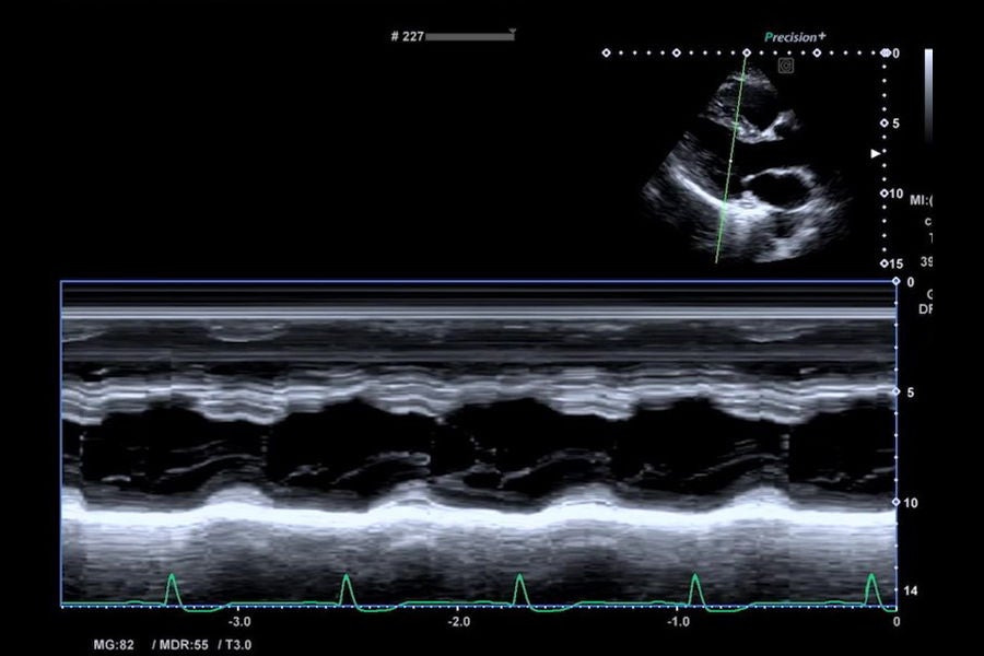
M for Cardiac
Watch Video
×
Section 6: Storing image data
Watch Video
×
Save images
Watch Video
×
Set the saved conditions
Watch Video
×
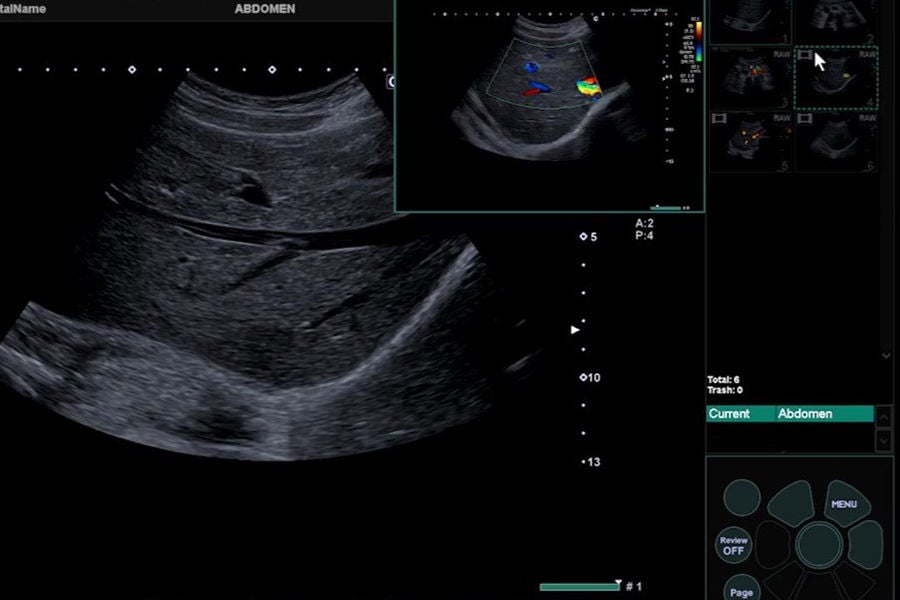
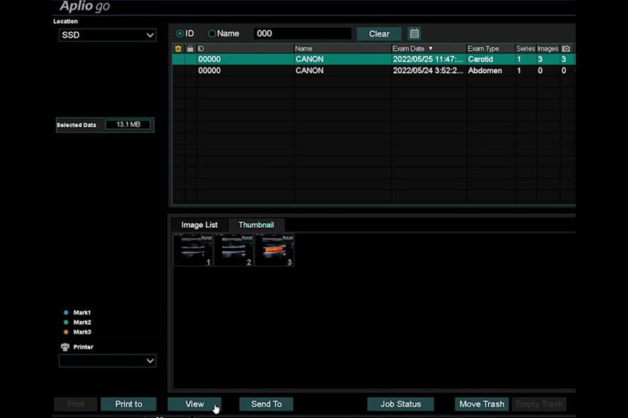
Review saved images
Watch Video
×
Section 7: Basic measurements
Watch Video
×
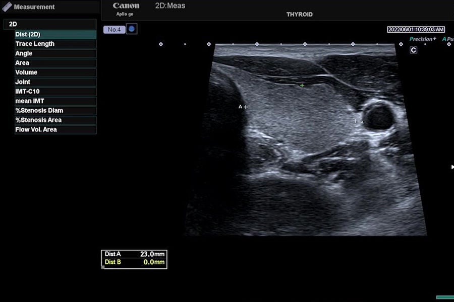
2D measurement
Watch Video
×
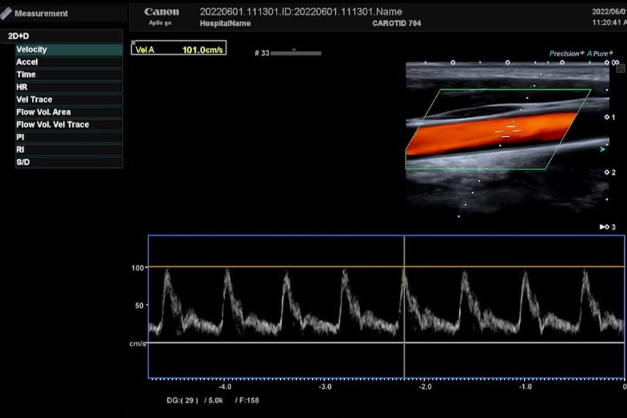
PW measurement(General Image)
Watch Video
×
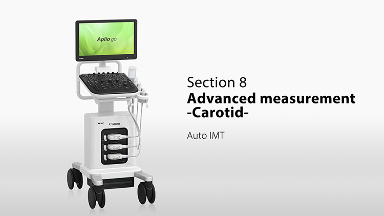
Section 8: Advanced measurements -Carotid-
Watch Video
×
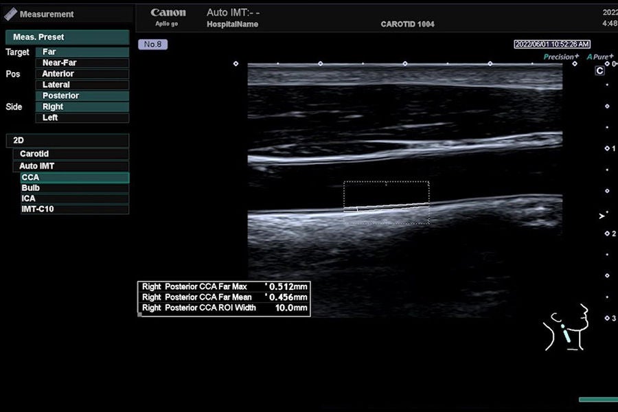
Auto IMT
Watch Video
×
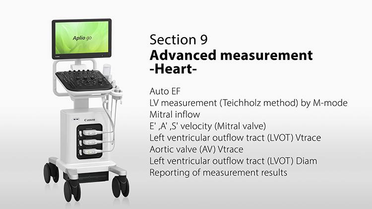
Section 9: Advanced measurements -Heart-
Watch Video
×
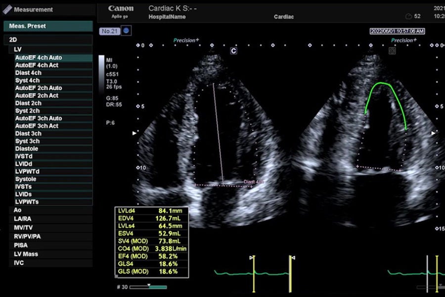
Auto EF
Watch Video
×
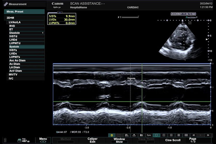
LV measurement (Teichholz method) by M-mode
Watch Video
×
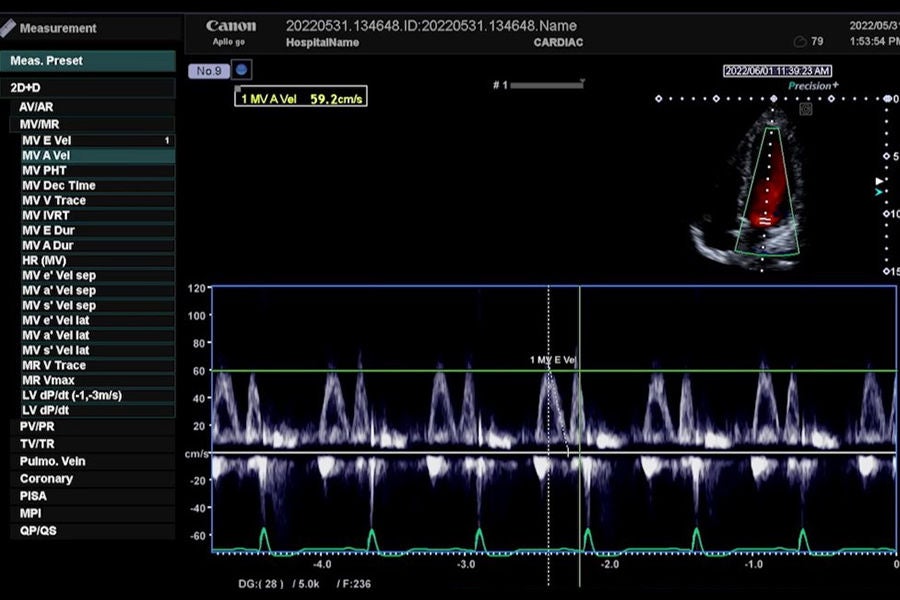
Mitral inflow
Watch Video
×
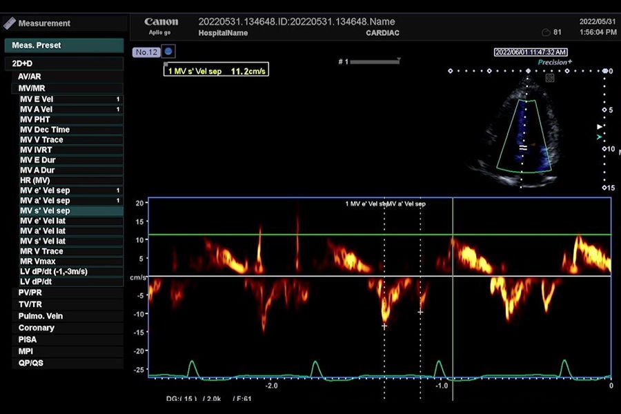
E' ,A' ,S' velocity (Mitral valve)
Watch Video
×
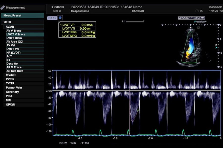
Left ventricular outflow tract (LVOT) Vtrace
Watch Video
×
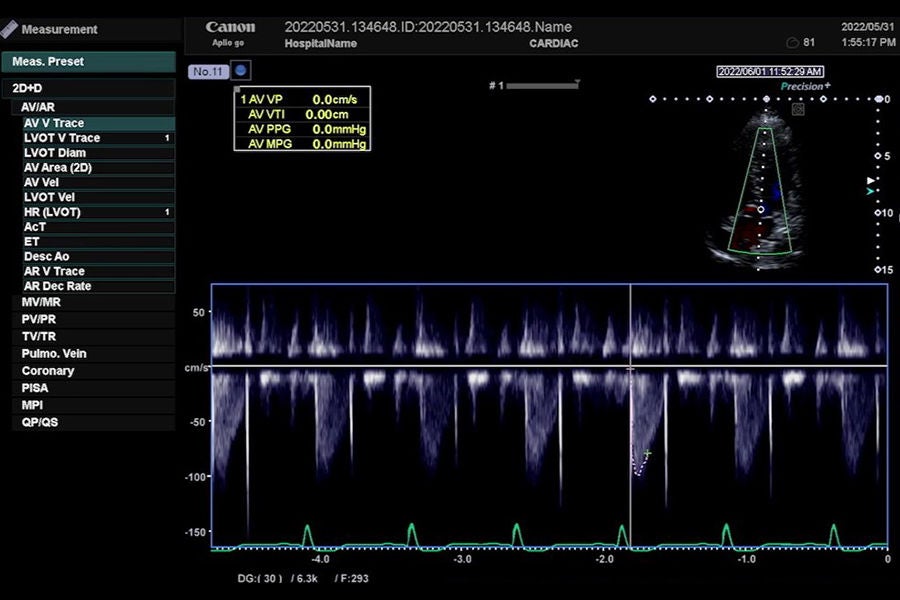
Aortic valve (AV) Vtrace
Watch Video
×
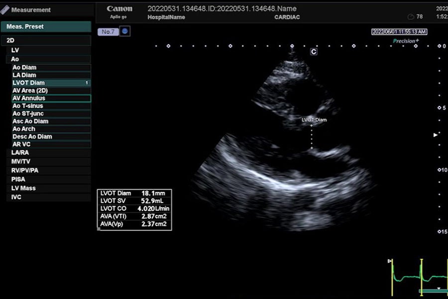
Left ventricular outflow tract (LVOT) Diam
Watch Video
×
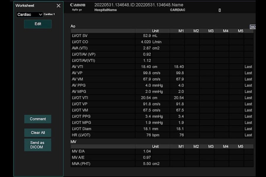
Reporting measurement results
Watch Video
×
Section 10: Advanced applications
Watch Video
×
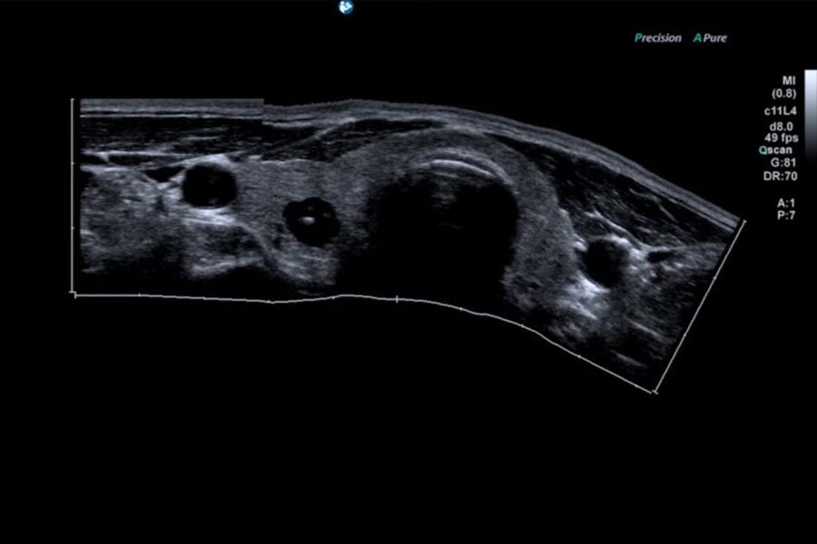
Panoramic View
Watch Video
×
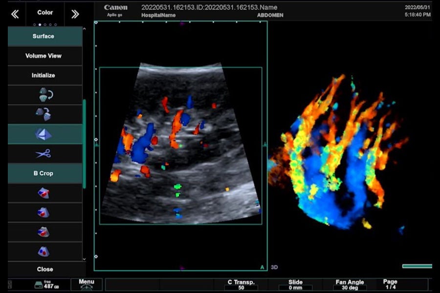
Smart 3D
Watch Video
×
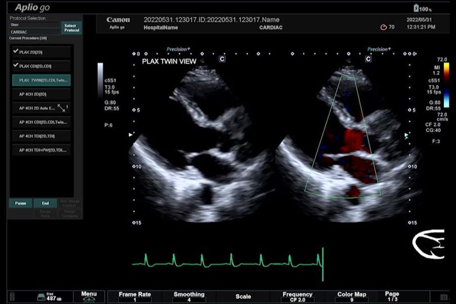
Protocol Assistant
Watch Video
×
Section 11: Patient browse
Watch Video
×
Display images
Watch Video
×
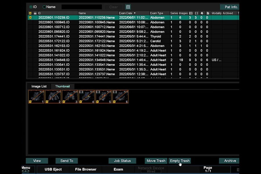
Delete images
Watch Video
×
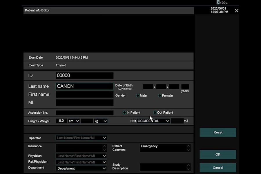
Change patient information
Watch Video
×
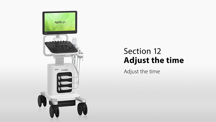
Section 12: Adjusting the time
Watch Video
×
Section 13: Shutdown
Watch Video
×
TOP
Product list
Ultrasound
lecture
Aplio go