

Case Study
Innovative High-Definition
Flat Panel Technology in the Treatment
of Cerebrovascular Disease
Adnan H. Siddiqui
MD, PhD, FACS, FAHA, FAANS Gates Vascular Institute
Kenneth V. Snyder
MD, PhD, FAANS Gates Vascular Institute
Betty Ashdown
Senior Clinical Marketing Manager ARRT(R), (CV) (CT), RCIS, FAVIR, FSICP
Canon Medical Systems, USA
Today more and more clinicians are performing endovascular stenting and coiling for treatment of intracranial aneurysms. Newer devices are being developed using materials that are more flexible to provide better durability, which allow the structural design of the devices to be smaller in size to improve patient outcomes.
Technology
Traditional flat panel detectors have difficulty in visualization of the newer devices being used today during minimally invasive interventional procedures. Digital Live Zoom can be used to increase image display size in real time during both fluoroscopy and digital acquisitions, from 1.2 to 2.4 times standard field of views (FOV: 12", 10", 8", or 6"). However using the highest live zoom modes tend to create digital distortion and pixilation in the image. Canon Medical’s new, technologically advanced High Definition detector technology boosts visualization and increases spatial resolution to facilitate the clinician in delivering safe patient care during interventional procedures.
History
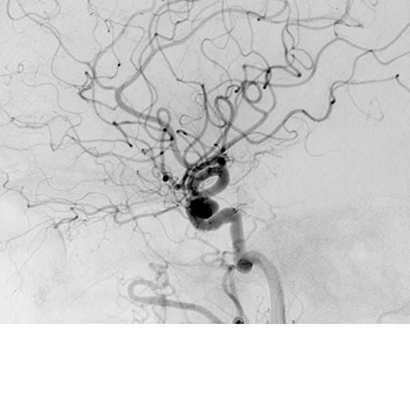
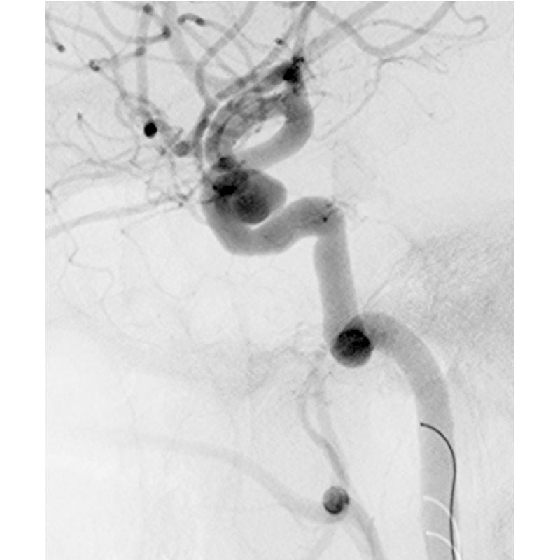
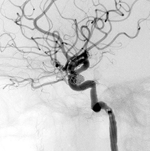
A 30-year-old female arrived at the emergency department complaining of the worst headache of her life. A CT scan of the head and a lumbar puncture were done, both were negative for the possibility of a subarachnoid hemorrhage. She then went for magnetic resonance angiography, which showed her to have an 11 mm right carotid cavernous sinus aneurysm.
Pre-Intervention Images
Procedure
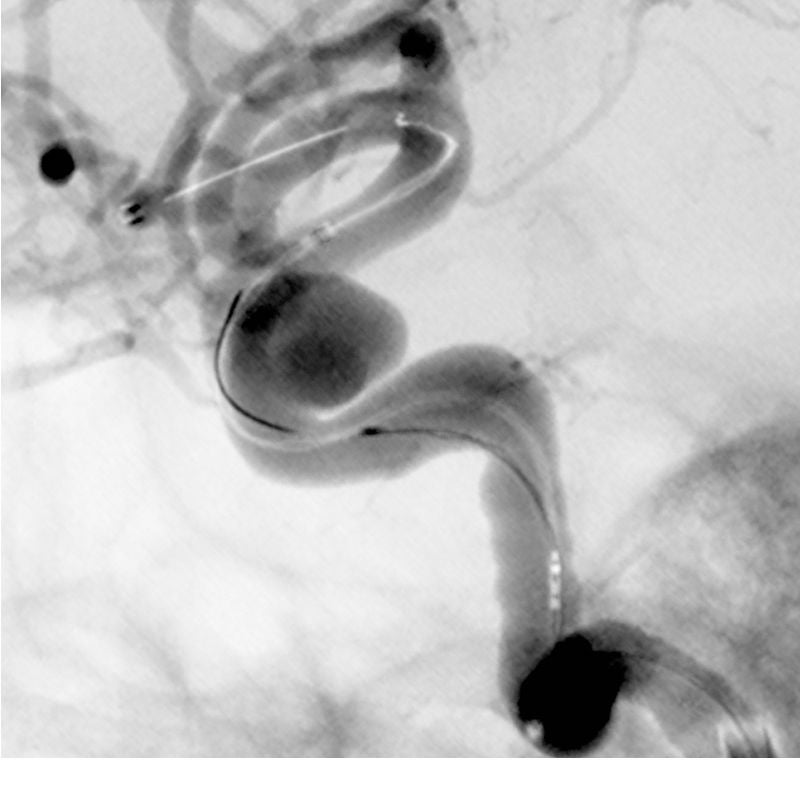
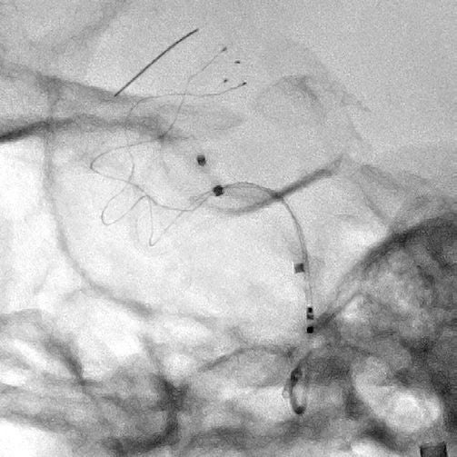
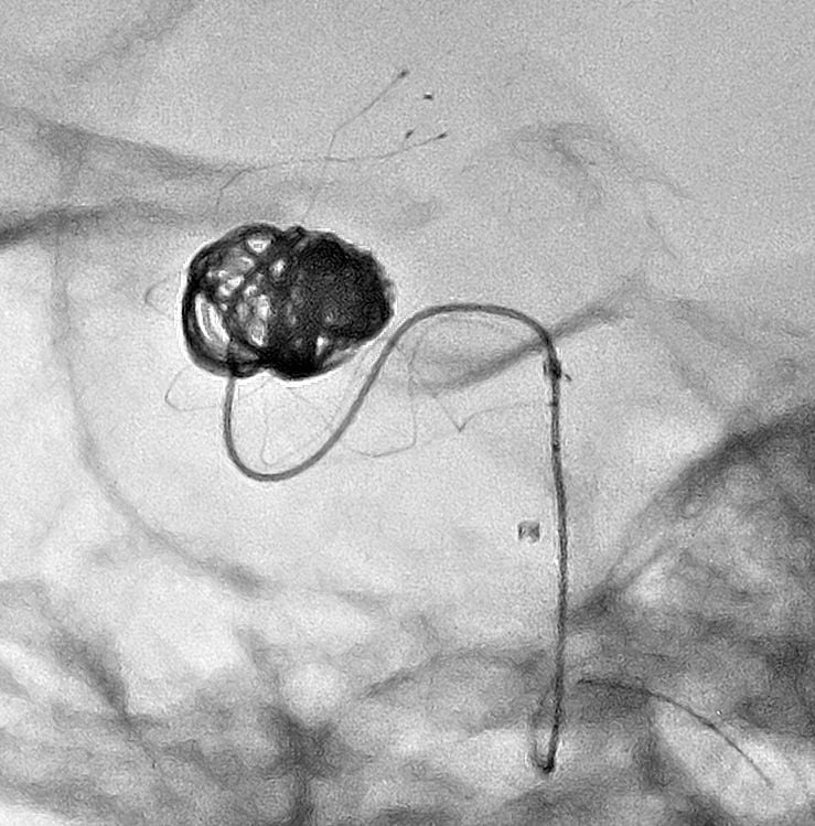
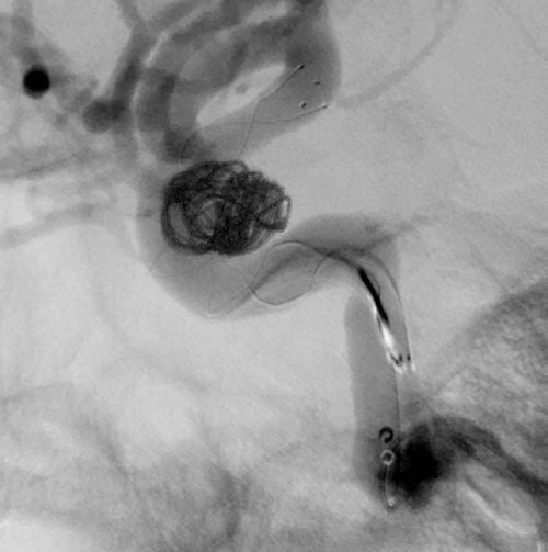
The physician decided to perform endovascular treatment of the aneurysm using a neurovascular stent and coils to prevent rupture. After femoral arterial access was obtained, a catheter was advanced to the right common carotid artery and digital subtraction angiography (DSA) was done. DSA imaging was used to obtain intra-arterial measurements of the carotid cavernous sinus aneurysm. The guidewire was then threaded through the carotid artery and a stent was positioned to cover the neck of the aneurysm. High-definition magnification modes of 3.0 and 2.3 inches were used to amplify visualization which aided in accurate insertion of the small coils through the stent struts and into the aneurysmal sac. Two long coils were necessary to pack the entire aneurysm volume.
Device Deployment Images
Post-Intervention Images
Conclusion
Canon Medical’s unique hybrid 12-inch flat panel detector design enables operators to maximize efficiency during interventional procedures and provides up to 2.5 times greater spatial resolution. Magnification modes of 3 inch, 2.3 inch, and 1.5 inch are now obtainable with remarkable resolution of the image. In this case, High Definition helped with stent positioning, ensuring the stent opened as expected, allowing for easy wire and catheter navigation through the stent tines, and allowed improved observation of coil deployment.
Images courtesy of Dr. Adnan Siddiqui at Gates Vascular Institute in Buffalo, NY
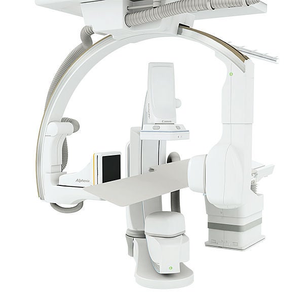
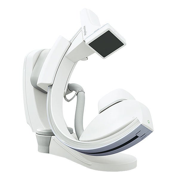
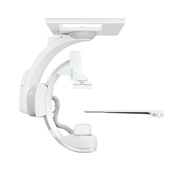
Related Angiography products
The content reflects information available at the time of publication and may differ from current information.
Social Media


