

Diving into the Future of CT with Deep Learning Spectral Imaging
Prof. Catherine Roy
trasbourg University Hospital is one of the top five research hospitals in France. It is renowned for the quality of its healthcare, clinical research and leadership in training. As a center of excellence in a wide range of specialties including cardiac surgery, transplantation, rheumatology, microsurgery, cryotherapy, immunology and surgical robotics, it contributes to advancing medicine globally. The hospital acquired an Aquilion ONE
/ PRISM Edition CT system from Canon in 2019.
Prof. Catherine Roy, head of the radiology department at the hospital, explains how its deep learning spectral imaging, powered by Altivity (the new AI brand from Canon Medical), delivers new diagnostic efficiencies.
The Aquilion ONE / PRISM Edition complements the Hospital’s existing Aquilion ONE / GENESIS Edition system, which was installed in 2018.
“With our vast and growing demand for high-quality imaging, our CT systems are at the heart of the hospital. They serve the needs of our numerous radiologists, clinicians and technologists,” said Prof. Roy. “Aquilion ONE / PRISM Edition has become invaluable for us, due to its reliability, fast
workflow and high versatility. In particular, it has impressed us through key technologies that deliver spectral imaging. The system is so useful that
my co-workers are almost fighting to work on the new machine!”
Spectral Imaging
“Featuring a single X-Ray tube with an automatic adaptive exposure, the field of view is up to a 50cm centi- meters, and scans be obtained by helical or sequential acquisition. Spectral reconstruction is performed by deep learning. With one click on the Vitrea workstation you can obtain a great
deal of information with the spectral analysis application. It's entirely automatic and doesn't take too much time. The Vitrea workstation
automatically provides you with a visceral and/or a vascular study. In addition, you can use spectral imaging in multiple body regions,” explained
Prof. Roy.
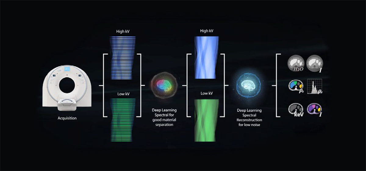 Deep learning spectral scanning, reconstruction and analysis.
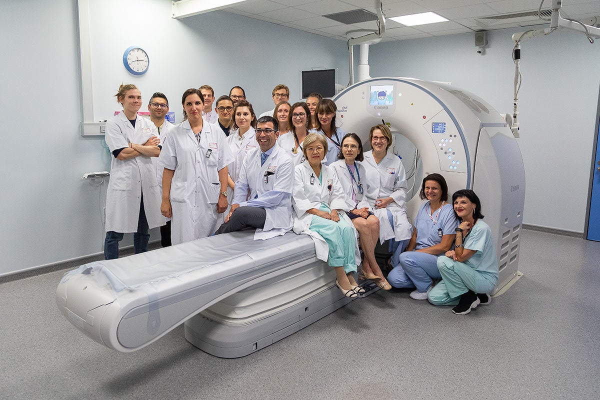 Radiology Team Strasbourg University Hospital, France.
 Monochromatic imaging from 35 KeV to 135 KeV showing improved contrast enhancement in the lower KeV images.
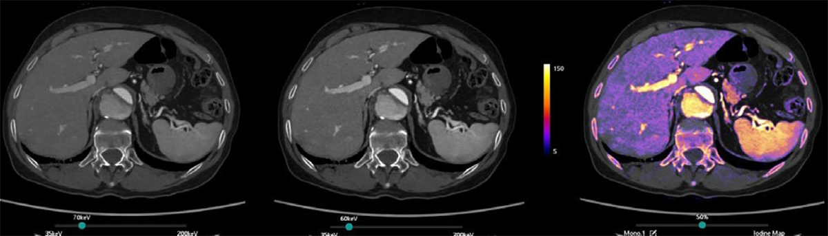 Despite the low iodine load of 100 ml of 270 mgI/ml and a flow of 1.3 ml/sec, Spectral CT helps to enhance the contrast at low keV free of noise. It provided an excellent contrast for the evaluation of the aortic dissection (type B).
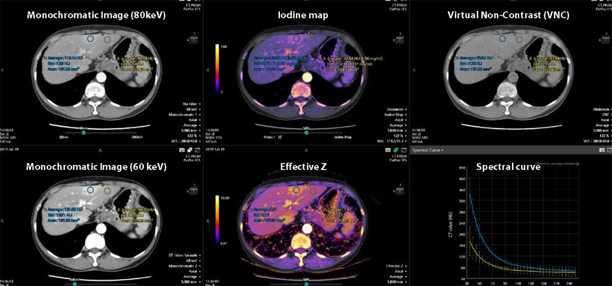 Vitrea Advanced Visualization Spectral analysis software.
Monochromatic images to see better
One type of image that is available with spectral imaging are high quality monochromatic images.
“We use the monochromatic images to better visualize to contrast on any abnormality. The monochromatic images produce excellent image quality
and to visualize iodine uptake even better, you can use a slidebar to adjust the keV level. Using the best contrast-to-nose ratio, you can optimize the
contrast inside the suspicious region by placing a region of interest,” she added.
“It's excellent for detecting abnormalities, even the small ones, and you can decrease the iodine loads, which is also very important. We use the
lowest volume or concentration of contrast agent possible. Concerning the radiation dose, it's quite similar to that obtained with iterative
reconstruction.”
Multiple tools to characterize and quantify
The Aquilion ONE / PRISM edition delivers a great deal of additional information that enables Prof. Roy and her team to better characterize and to
quantify what they are scanning.
“You obtain an iodine map, which is the information on the iodine uptake; the virtual non-contrast (VNC), with no iodine at all; and you can select
an evaluation of the spectral curve by placing a region of interest and evaluation of the effective Z image or electron density image. With these
three elements, you can differentiate contrast enhancement from non-contrast enhancement. With the VNC, you can avoid the non-contrast acquisition that was previously done,” she continued.
“Supplementary information available with the software is to have the quantification of the iodine load in mg per ml. You can obtain the spectral
curve by placing a region of interest inside the tissue.”
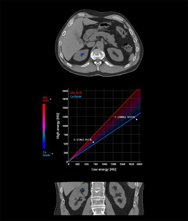
“Additionally, iodine maps improve visibility of nodules with iodine uptake. For examinations, like scanning for kidney stones, the software is very
easy to use. In the future, I guess that we will have a better evaluation of tissue perfusion, for instance: for the follow-up of oncologic patients and to
prove that there is an efficiency of antiangiogenic treatment. Or perhaps for immunotherapy. With just only one injected phase, you can
characterize tissue.”
Extraordinary Images
The Aquilion ONE / PRISM Edition, with Deep Learning Spectral technology powered by Altivity, has been able to meet the needs of the whole team
at the Hospital.
“The best descriptive classification of the Aquilion ONE /
PRISM Edition CT is - Versatility for all diagnostic purposes.”
Prof. Catherine Roy, Head of the Radiology department at the Strasbourg University Hospital, France.
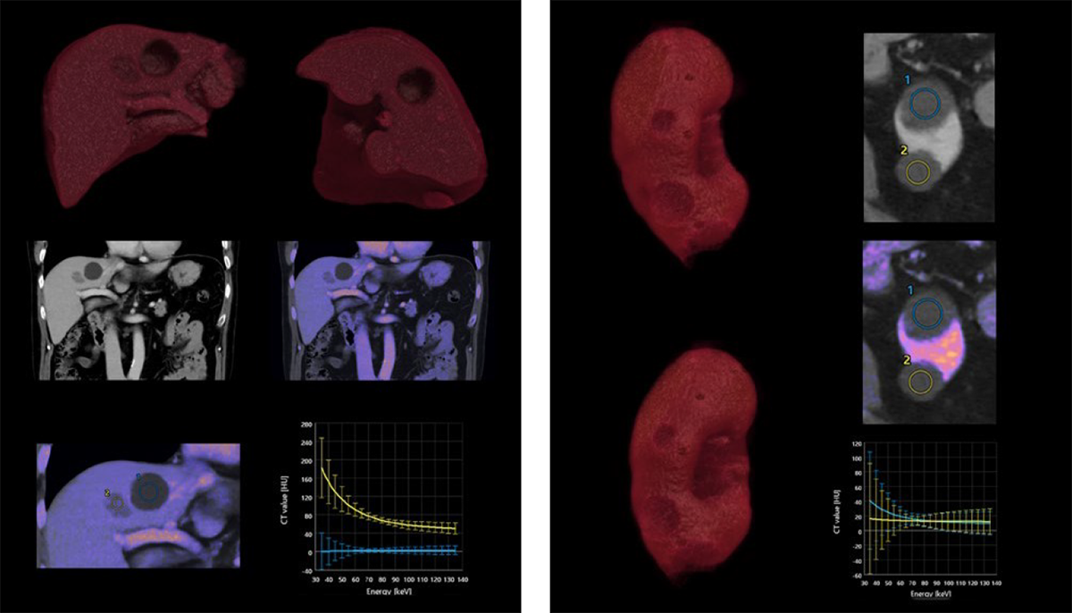 Deep Learning Spectral CT used to evaluate liver and renal masses. Spectral DLR delivers excellent energy separation, enabling assessment of Iodine uptake. Additionally, the Iodine map clearly outlines the regions of Iodine uptake.
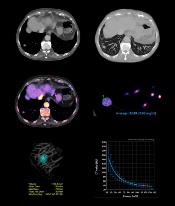
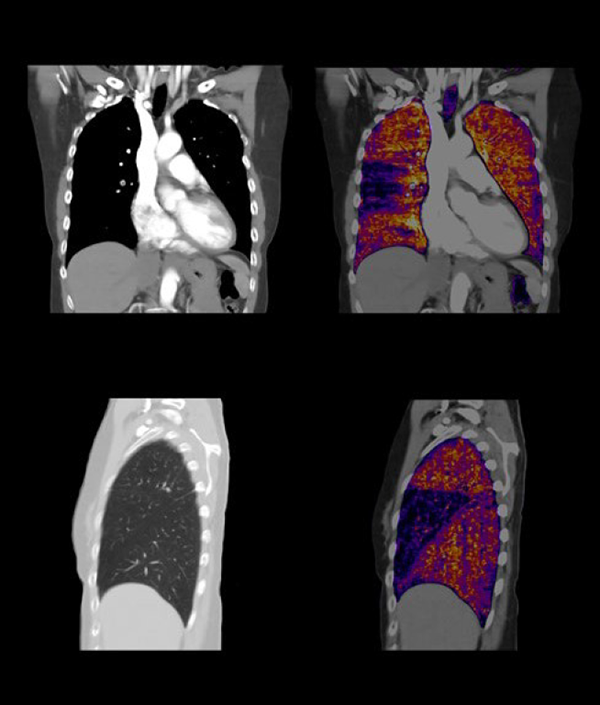
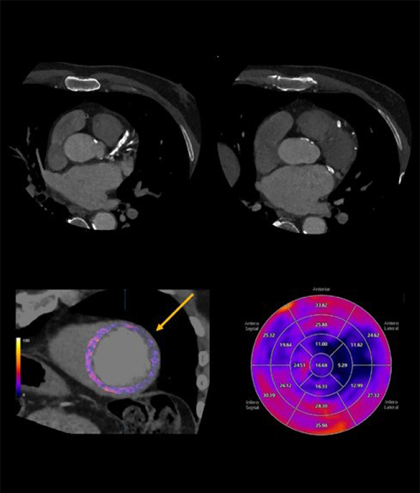
“Our clinicians love, and they are very interested by innovative things, especially with spectral imaging. Their feeling is that with this new scanner's
spectral imaging capabilities, they are more confident with their diagnoses,” said Prof. Roy.
“Our technologists need easy-to-use hardware and software and this is available with the Aquilion ONE / PRISM Edition. They need a reliable
scanner, and this is especially true for the Aquilion ONE / PRISM Edition. There have been no problems with it at all. The best descriptive
classification of the Aquilion ONE / PRISM Edition CT is - Versatility for all diagnosis purposes.” //
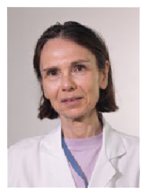
Prof. Catherine Roy
Head of the Radiology department at the Strasbourg University
DISCLAIMER: The clinical results described in this paper are the experience of the author. Results may vary due to clinical setting, patient
presentation and other factors.
The uses discussed in this paper may not have Regulatory Approvals in all regions, please discuss with your local sales company.
The content reflects information available at the time of publication and may differ from current information.
Social Media


