

Canon Medical in Iceland: Landspitali Experiences
 Photography: by Cojan van Toor, VISIONS professional photographer. Photo: Iceland Place: Gullfoss Falls (southwest Iceland)
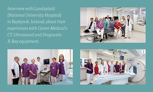
Pétur Hannesson and Steinuum Erla Thoracius, both Head of the Radiology department, and Kristín Rut Haraldóttir, responsible for the education and training of midwives in Ultrasound at Landspitali (National University Hospital) in Reykjavik, Iceland, share their experiences with Canon Medical’s equipment in this dedicated interview with VISIONS magazine.
With 636 beds, Land Hospital in Reykjavik is the leading hospital in Iceland and the largest workplace for healthcare professionals. The facility is divided in two buildings and provides the country with every kind of care and imaging procedures. One of the challenges is to maintain radiology equipment on both sides, according to Pétur Hannesson, Head of the Radiology department.
“We have everything under one roof. We’re a medium sized hospital but we attend a population of 360,000 people, so it’s also challenging for our team. We’re both a primary hospital and a tertiary hospital. Everything is done here. We deal with all cases and all situations,” he said.

“The Aquilion ONE / GENESIS Edition is equipped with a 16 cm detector, which makes it an ideal scanner for cardiac and angiography examinations.”
Pétur Hannesson, Head of Radiology at Land Hospital. He studied medicine in Iceland and received his medical degree from the University of Iceland. He did his specialization in radiology in Lund, and started working at Landspítali in 1995. His subspecialty is oncology.
CT imaging
The latest CT scanner at the hospital is the Aquilion ONE / GENESIS Edition. The staff are very happy with the system, especially with the speed at which it can help to perform examinations, according to Pétur Hanneson.
“The Aquilion ONE / GENESIS Edition is equipped with a 16 cm detector, which makes it an ideal scanner for cardiac and angiography examinations. With this system it is possible to scan the heart in a single rotation or one heartbeat. The wide volume coverage of the detector also gives us the opportunity to scan paediatric patients extremely fast and without sedation. We’re very pleased with the capabilities of the system,” he said.
Pétur Hanneson and his team are carrying out an increasing number of cardiac examinations every week, reducing interventional examinations and traditional angiography for diagnostic purposes. “CT is a wonderful tool, a very strong ally in that regard,” he said.
The capacity to perform coronary studies was key in the decision to select Canon Medical equipment, according to Steinuum Erla Thoracius, Head of the Radiography department.
“We wanted a good system for coronary imaging. The large area detector of the Aquilion ONE / GENESIS Edition allows us the scan the heart very fast with only one rotation. This results in lower radiation doses for the patients and administering less contrast media. I liked that part very much,” she said.
Paediatric examinations are rare in our hospital, but the Aquilion ONE / GENESIS Edition is also helpful in this area, thanks to its ability to lower the dose whilst increasing acquisition speed. The latest iterative reconstruction technique, which lowers the noise in the images and improves image quality, which is very important.
“Dose has been going down for the last years and that’s why it’s important to have new equipment, to enjoy the latest advances in dose reduction,” Pétur Hanneson said.
The Aquilion PRIME is the department’s workhorse and is used for almost everything, particularly trauma imaging. “It’s very important for trauma centres to have two CT scanners. Imagine not to be able to scan trauma patients because a scanner is in use or undergoing regular maintenance. We must be able to scan trauma patients at any time,” Pétur Hanneson said.
The Aquilion PRIME comes with a series of benefits, starting with the “insta-view”, a beloved option among radiographers, according to Steinuum Erla Thoracius. “Insta-view”allows us to review the images immediately while the scan is made. “
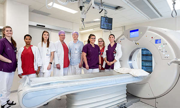
“A valuable asset is the user friendliness of the Aquilion PRIME.”
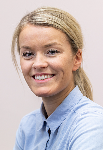
Steinuum Erla Thoracius, Head of the department of Radiography at Landspítali, where she has been working for over ten years. She has a bachelor and diploma degree in Radiography and a Masters’ degree in Human Resource Management.
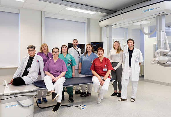
“The Adora DRFi is good for high quality skeletal examinations and it’s also easy to move.”
Diagnostic X-ray: the Adora DRFi
Fluoroscopy is no longer a popular trend and most fluoroscopy units in Scandinavia are waiting for an occasional examination. Petur Hanneson once had four fluoroscopy units in his department, but now offers a lot of examinations using barium swallow contrast material as an alternative.
“You don’t do colon x-ray examinations anymore, but either go for endoscopy or CT. Things have changed a lot,” Pétur Hannesson said.
Still, it is important for radiology services to continue to offer fluoroscopy, and this is why Hannesson and Thoracius chose the Adora DRFi system.
“We use the equipment 95% of the time for normal x-ray, but the possibility to use it also for fluoroscopy was very appealing. We use the space for two purposes and don’t need an extra fluoroscopy room, so having the Adora DRFi made a lot of sense,” Pétur Hannesson said.
Image quality is excellent with this system as well, and the system is very user friendly, according to Thoracius.
“The Adora DRFi is good for high quality skeletal examinations. It’s also easy to move, which is very important for radiographers. It has good ergonomics. It’s not a mobile x-ray unit, but it’s really easy to move the x-ray tube,” she said.
Landspítali radiographers’ input is always key in the decision to purchase new equipment and ergonomics are always a strong selling point. “They have a unique point of view, as they work with the machine all day. The person who is going to use the equipment knows better what to expect and what matters,” she said.
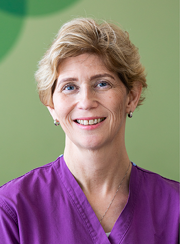
“The Aplio 400 has light probes, delivers good image quality and works very well.”
Kristín Rut Haraldóttir, specialised midwife at Landspitali. She graduated as a nurse in 1983 and became a midwife referee in 1985. She completed her Masters’ degree in 2012. She learned how to use ultrasound in 1988 and has been using the modality exclusively since 1995.
A midwife’s experience with Ultrasound
Kristín Rut Haraldóttir is responsible for the education and training of midwives in ultrasound at Landspitali. Teaching is crucial as midwives perform 95% of all fetal ultrasound examinations in Iceland. Landspitali’s Prenatal Department has eight midwives and three doctors who are specialised in prenatal screening. The team carries out around 11,000 fetal scans per year, from the typical 12 to 20 weeks and growth scans to Doppler and invasive tests like amniocentesis and chorion biopsy.
“Every year we can expect to pick up 60 to 100 fetal abnormalities, so it’s very important to have qualified staff and good equipment. You can’t do precise fetal examinations without good equipment,” she said.
In 2016 the hospital bought three Aplio 400 systems to replace the 11-year-old GE equipment. Workflow has improved tremendously ever since.
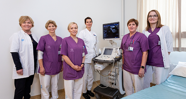
Since we do 11,000 scans per year, we need something that’s easy and works,” she said. Kristín Rut Haraldóttir carries out typical examinations with the Aplio. The Advanced Dynamic Flow (ADF) Doppler is extremely useful to depict not just the heart, but also other organs. “We can see the heart extremely well to pick up big defects early on. You have a clear signal and clear image of the heart. I can also see the stomach and the bladder, and I can measure the risk for Down syndrome. It’s truly amazing.”
Measurements can be done in a row: the fetal head circumference, the leg, etc. “Canon makes it so easy for us. You just enter next, next, next and next, and you can do all your measurements,” she said.
The equipment is user friendly and fits everyone’s size. “You can turn and move the system very easily. All of us can use it: I’m 1m60 and the next midwife is very tall, so you don’t lose time adjusting the equipment.”
Low noise is a definite plus. “You don’t hear the machine when it’s on. When you work all day in a small room and there’s a lot noise from the system that can really be annoying. With the Aplio, that doesn’t happen. There’s been a big improvement in the last years,” she said.
The equipment is easy to clean, another benefit when gel has to be used repeatedly. “The keyboard is easy to access, as it is just under the console, which is very spacious. There’s no problem with gel. If it falls, you can clean the board easily. That’s very important. They have thought about everything. You just need to press a few buttons.”
Cooperation with Canon Medical has been crucial in training staff. “It’s tailor made. We lose less time with the machine and can spend more time with the patient. A member from Canon Medical was with us for a few days to teach us everything. I can phone her if I have a problem,” she said. //
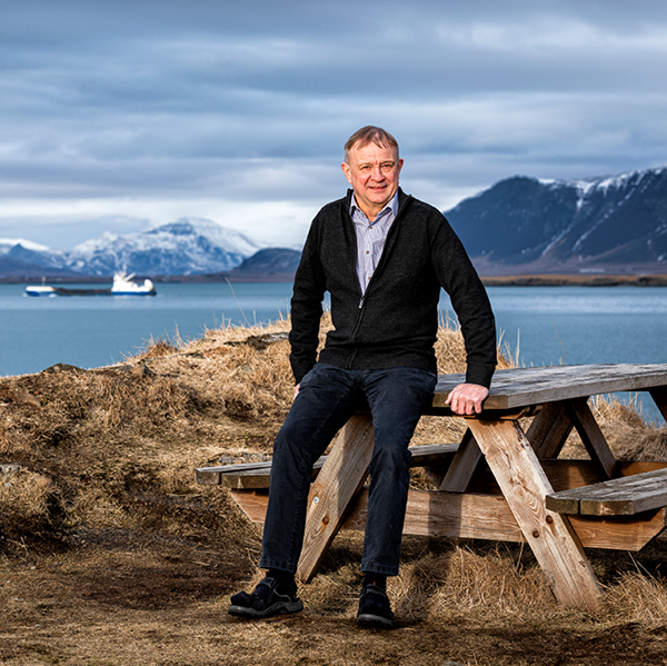
“It’s always a pleasure to work with Canon Medical Systems Europe. The communication is always very good, which is important for our direct communication with the customers in Iceland.”
Hlöðver Þorsteinsson, Director of RL hf. – Rafland (Canon Medical‘s Partner in Iceland)
Canon Medical’s Partner in Iceland - RL hf. – Rafland
RL hf. - Rafland was established 1964 and has always been a family company. The cooperation with Canon Medical (formerly Toshiba Medical) started in 1970 but at that time only selling Toshiba CTVs, HiFis and later also microwave ovens. Also at that time the company was in cooperation with Canon Corporation for project for turbines for the first Hydropower station in Iceland.
In 1985 the company started cooperation with Canon Medical where the first CT in Iceland was sold to Landspitali in 1986. Through the years the company has been working close to the healthcare system, both private and governmental and always with excellent and trustworthy support from the team of Canon Medical Systems Europe. Canon Medical is known for their reliability and for excellent service. Together with Canon Medical, RL hf. - Rafland has become one of the leading companies, with the highest market share in X-ray business, in Iceland.
The content reflects information available at the time of publication and may differ from current information.
Social Media


