

High-Resolution Sonography Redefines Hand Surgery Diagnostics
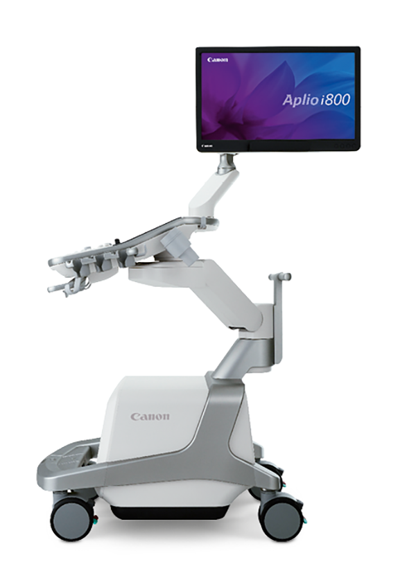
The Handchirurgie Seefeld (hand surgery) clinic in Zürich, Switzerland, relies on high-tech medicine with the state-of-the-art Aplio i800 ultrasound diagnostic system of Canon Medical.
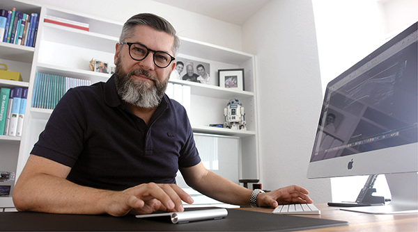 Dr. Sebastian Kluge, FMH professional association specialist doctor for surgery and hand surgery, ultrasound diagnostics, musculoskeletal system SGUM (Swiss Society of Ultrasound in Medicine), appreciates the huge advantages of sonography for hand surgery because it provides insights like no other diagnostic method and therefore redefines the causes of diagnosis.
The major challenge in hand surgery is the precise handling of delicate structures. There is hardly any other specialty where elements of plastic surgery, orthopaedic surgery and micro surgery are so closely related and deal with extremely complex and small anatomical structures. Therefore, diagnosis with sub-millimetre-level accuracy has a significant impact on optimal treatment success. Specialists like Dr. Sebastian Kluge, FMH professional association specialist doctor for surgery and hand surgery, ultrasound diagnostics, musculoskeletal system SGUM (Swiss Society of Ultrasound in Medicine), use the high-tech Aplio i800 sonography device to redefine the approach to a highly differentiated field. “The Aplio presents structures and situations in a spatial resolution which would not be possible for any CT or MRI, whilst simultaneously providing the possibility of dynamic assessment,” he explains.
Dr. Sebastian Kluge started his medical career around 20 years ago at the Berufsgenossenschaftliche Unfallklinik (occupational accident clinic) in Ludwigshafen, where he completed civilian service after finishing school and discovered his interest in hand surgery. “Until that point I was all set on studying drums and piano. It was only my fascination with hand surgery that made me embark on a career in medicine.” Today he is one of the most well-known specialists in his field, and his expertise makes him highly respected by his patients and his medical colleagues alike. He has delivered lectures and ultrasound seminars around the world and worked with fellow specialists to write a standard textbook entitled “Ultraschalldiagnostik der Hand” (Diagnostic Ultrasound of the hand).
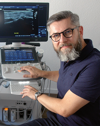
“The presentation of soft tissue and bone surface and their dynamic interaction with a resolution of 24 MHz is staggering and proves the huge potential of sonography.”
Dr. Sebastian Kluge, FMH professional association specialist doctor for surgery and hand surgery, ultrasound diagnostics, musculoskeletal system SGUM (Swiss Society of Ultrasound in Medicine).
Ultra-high frequency redefines causes of diagnoses
The Aplio i800 sonography device from Canon Medical is an important “hand tool” in his daily routine. He decided to invest because “this system offers previously unforeseen new possibilities in hand surgery.” By this he means in particular the extremely high resolution that is possible in combination with the 24 MHz probe, therefore enabling the differentiation of structures down to a sub-millimetre level. In particular, where the small distance between the skin surface and bone means that a high penetration depth is not needed, the system displays a sonographic image in the highest resolution. Dr. Kluge therefore sees big advantages for high-frequency ultrasound in rheumatology, neurology and orthopaedics.
Dr. Kluge’s specialties are post-traumatic changes in the forearm, wrist and fingers, the non-operative and operative treatment of nerve compression syndromes and inflammatory-degenerative and arthrotic changes in the hand and finger joints, including joint replacement. “The spectrum of causes problems and the range of treatment options available are immense”, the doctor explains. “In this respect, various suspected diagnoses need to be isolated from each other extremely precisely and treatments need to be planned and implemented with the utmost precision,” he added.
The Aplio i800 is ideally suited for all of this work. This is because while sonography was previously carried out at sound frequencies of between 15 MHz and 18 MHz, Canon Medical offers matrix technology in the ultra-high frequencies of 24 MHz and 33 MHz. “The resulting increase in spatial resolution not only allows the person carrying out the examination to take the very important step of moving from a suspected diagnosis to absolute diagnostic certainty for many matters, it actually redefines the cause of certain diagnoses.”
High-resolution imaging allows for high-precision interventions
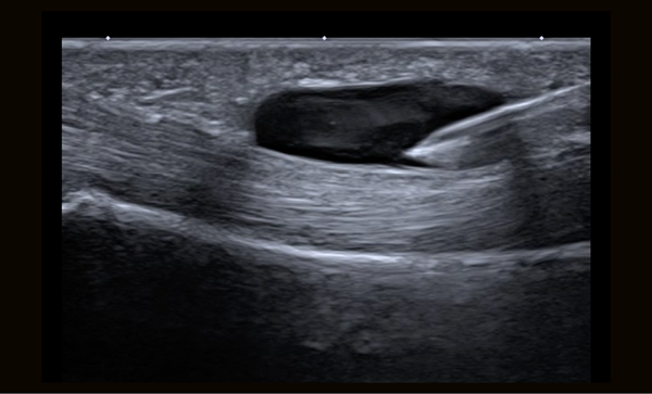
The extremely high resolution of the sonography system provides valuable support even for ultrasound-controlled interventions. The most common indication in hand surgery is stenosing tenosynovitis. This imbalance between the annular pulley width and flexor tendon thickness may result in clicking of the affected finger. When attempting non-operative treatment, a cortisone injection can even be given with ultrasound support. This results in a lower rate of side effects due to more precise placement of the needle in the digital canal. However, the method only really comes into play in the event of differentiated hand surgery interventions such as occult wrist ganglions, which are “destroyed” under sonographic guidance, so in many cases the complaints can be permanently eliminated even without surgery. “In these special cases, the immense spatial resolution even allows for targeted ultrasound-controlled anaesthesia of the nerve supplying the wrist joint capsule, which makes this a pain-free intervention. Even minimally-invasive carpal tunnel operations can now be done under sonographic guidance.”
Another example for the usefulness of high-resolution ultrasound can be seen in the case of post-operative complications. Implant protrusions, which may occur occasionally after surgery on radial fractures, for example, may cause irritation of adjacent flexor and extensor tendons. This often results in an extensor tendon rupture, usually of the long extensor tendon in the thumb. “The ideal method for localising any implant-related complications of this kind is ultrasound”, says Dr. Kluge. In these cases, CT shows the implant protrusion but not the soft tissue trauma caused, while in an MR scan the soft tissue injury is frequently covered by metal artefacts. “In this case, sonography allows for optimum differentiation of bone surface, soft tissue and implant – in high resolution”, Dr. Kluge summarised.
“We not only have a responsibility to the patient, but also to our colleagues.” Patients are also repeatedly referred by fellow hand surgeons for ultrasound diagnosis – usually with very precise and differentiated problems. “In this case in particular, the expectations of a precise diagnosis are extremely high, because our colleagues base their further treatment on this diagnosis”, said Dr. Kluge.
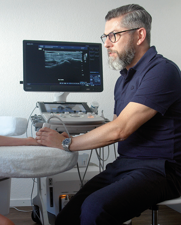 In combination with the 24 MHz ultrasound probe, structures can be shown precisely and without artefacts.
Optimal ease-of-use makes work processes easier
The hand specialist has got to know the advantages of ultrasound technology “from the bottom up”. In the wards where he completed his medical training in Frankfurt, Bern and Zurich, great importance was always placed on sonography. This is why he gives lectures and seminars on diagnostic and treatment options with the aid of modern sonography systems.
Dr. Kluge gets to the heart of the matter when he says “the processing of the sound information and the resulting image quality in the new Canon Aplio systems are phenomenal.” They can be used to produce very exact findings. Even for the finest structures such as digital nerves it is possible to differentiate between intact and destroyed nerve fibres. This is particularly helpful when planning nerve reconstructions, because the length and thickness of a nerve transplant which is needed can be precisely measured before the operation and nerve transplants can be planned or ordered in the correct size. In his experience, “the Aplio i800 is also an extremely user-friendly system”. An example of this is “when the program is selected, the system also changes to an optimised key assignment in the control panel, which means that there is no need to search for functions unnecessarily.”
However, the perfect integration of the device into the practice’s work processes is also important for Dr. Kluge. The ultrasound device is integrated into the practice’s information system via a wireless network. After the hand specialist has carried out a clinical examination on a patient, he can access all of the patient information necessary for the examination on the Aplio i800 and then connect further diagnostics there immediately. The results are then automatically transferred to the practice’s software and are available there for further processing.
“An accelerated workflow is just as important for us as comprehensive clinical examination is for our patients. Suspected clinical diagnosis are often able to be confirmed or excluded during the initial consultation thanks to the sonography.
In many cases, there is absolutely no need for additional imaging procedures such as CT and MR, which in the latter case also results in cost reductions and reduced radiation for the patient from CT”, the hand specialist explains. Thanks to the iDMS (intelligent Dynamic Micro-Slice) function of the matrix probes from Canon Medical, the crystal elements can be aligned so that a very narrow and focus-independent sound field is produced. “This “sharpening (of) imaging slice thickness” makes it possible to focus on even the smallest joint spaces between individual carpal bones”, stresses Dr. Kluge.
Sonography will remain exciting in future
Sonographic imaging has played a key role in the life of doctor and passionate music lover Sebastian Kluge for around 20 years, and to some extent he thinks of it as a work of art. He has been using it for a good 15 years as a hand surgeon and knows that “this diagnostic technology offers enormous opportunities that have by no means been exhausted yet”. He looks forward to the next developments, which he is monitoring keenly. He has already been able to scrutinise the Aplio i800 in advance as part of his own advanced training events.
He thinks there is great potential in the further definition of software algorithms and image processing, and also in the further miniaturisation of the systems. He said “making high tech ultrasound systems like the Aplio i800 from Canon Medical portable would be a vision that would be worth achieving in many respects, not least because it would mean that high-frequency imaging could even be used in ultrasound-guided operations on the hand without having to be tied to a single operation site.” //
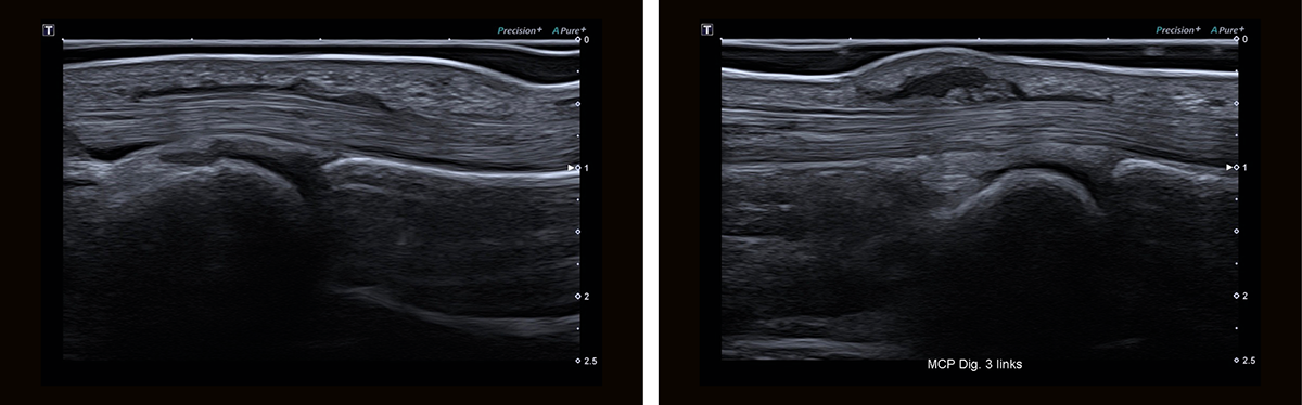
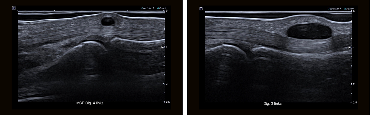
Stenosing tenosynovitis. This is one of the most common conditions in the hand. In addition to primary causes such as the thickening of the A1 annular tendon at the level of the joint at the base of the finger (left), the condition can also be caused by synovitis of the flexor tendons. Subcutaneous Dupuytren's nodules can also radiate into the digital canal or the A1 annular pulley and result in a secondary constriction of the digital canal (right). A differentiation, which is relevant for the therapeutic approach, can be made reliably using sonography.
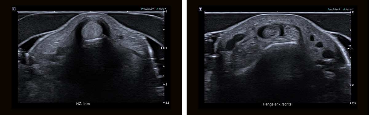
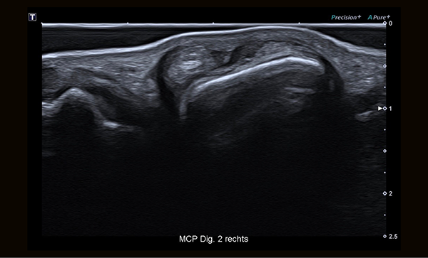
De Quervain's tenosynovitis. This is an entrapment syndrome of the first extensor tendon compartment. The tendons of the abductor pollicis longus and extensor pollicis brevis muscles are compromised and can barely be differentiated from each other as individual structures anymore, while the extensor retinaculum displays a halo-like thickening (left). Some patients present with an additional partition of the first extensor tendon compartment (subsheath), which can also be easily distinguished using sonography (right). Knowledge of this is important for treatment, as patients with a subsheath do not respond as well to non-surgical treatments.
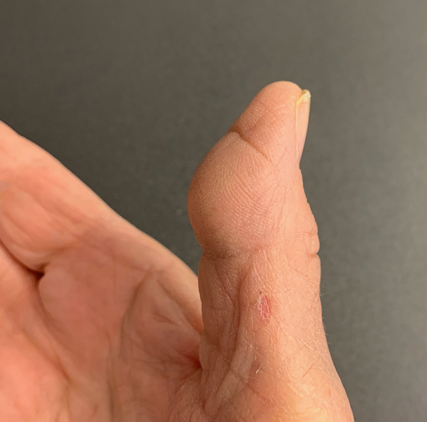
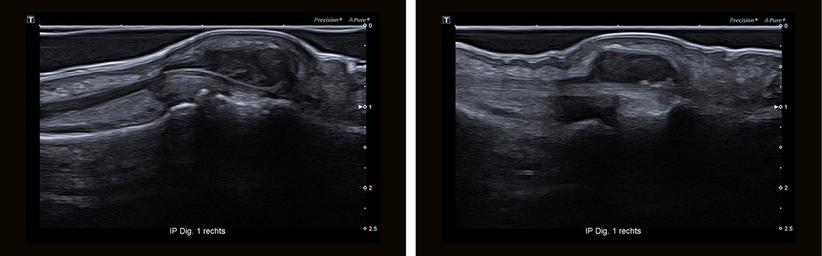
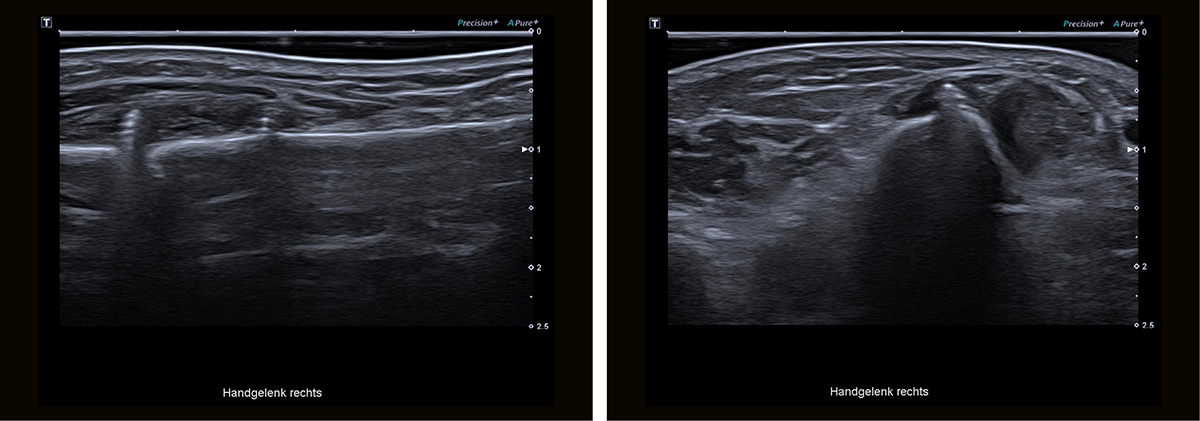
Implant-related complications. Postoperative complications caused by osteosynthesis materials which have been inserted are not uncommon. In the case of radial fractures treated using surgery in particular, prominent plate designs or excess screw length may result in irritation and a rupture of the flexor and extensor tendons. In this situation, the diagnostic advantage of sonography in comparison with CT relates to the simultaneous presentation of the cortex, relevant implant protrusion and soft issue, and the advantage over MRI relates to the absence of metal artefacts.
The content reflects information available at the time of publication and may differ from current information.
Social Media


